Tibial Head Fracture
What is a Tibial Head Fracture?
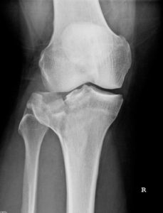
A tibial head fracture is defined as a lower leg fracture in the knee joint area. This can occur as a split, comminuted, or impression fracture and lead to a complete defect of one or both sides of the knee joint.
How is a Tibial Head Fracture Treated?

Tibial head fracture
In any case, in addition to an X-ray examination, a computed tomography (CT) scan should be performed to accurately assess the joint surface. If a fracture causes step-offs of more than 2 mm in the weight-bearing zone, these should be corrected surgically. This prevents future pain. Joint function is restored, and the operation also serves to prevent post-traumatic osteoarthritis.
During the operation, the fracture is manipulated into the correct position, with the position of the joint fragments being controlled arthroscopically, meaning under direct magnified view using a rod optic within the joint.
The corrected position of the fracture is held in place either by screws only, or also by a titanium plate.
During arthroscopy, associated injuries are also diagnosed and treated immediately: meniscus tears and cruciate ligament tears are common associated injuries with tibial head fractures, which can usually be primarily reconstructed by refixation or suturing.
Do I Need Physiotherapy after the Operation?
Early functional rehabilitation is initiated post-operatively. You can begin physiotherapy and movement exercises immediately after the operation.
Alternative Treatment Methods for Tibial Head Fracture
Alternatively, the tibial head fracture can be treated with a splint or cast. In this case, immobilization must be maintained for 8-12 weeks. Only then, after removal, can mobilization of the knee joint begin.
Supracondylar Fractures

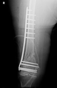
Supracondylar Fracture Surgery
If a fall causes a fracture just above the knee joint, this is referred to as a supracondylar fracture. This type of fracture is usually accompanied by fragment displacement due to muscle pull, and is therefore in most cases surgically treated with an angle-stable titanium plate or an intramedullary nail inserted from the knee joint side.
These fractures can also be treated with early functional rehabilitation after a correctly performed operation.
Patellar Fracture – Patellar Fracture
What is a patellar fracture?
The kneecap (patella) is part of the extensor mechanism of the knee. A patellar fracture is a fracture of the kneecap.
What are the causes of a patellar fracture?
The cause of a patellar fracture is usually direct force. The typical accident mechanism is a fall onto the knee. Often, a hard edge is involved (stair step, curb), which the kneecap hits. After a patellar fracture, the bone fragments are no longer in the correct position.

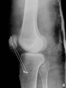
Patellar tendon rupture
How is a patellar fracture treated?
Only in rare cases can a patellar fracture be treated conservatively.
The operation is performed openly. The procedure depends on how the kneecap was fractured. Fractures with displaced fragments must first be brought back into the correct alignment. Especially on the back of the kneecap, a smooth result must be achieved.
Longitudinal fractures can be screwed in from the side.
Transverse fractures are usually held in place with two wires passed longitudinally through the fragments. The wires are tightened by another wire (tension banding). Through this tightening, the fragments grow back together.
Do I need physiotherapy after the operation?
Immediately after the operation, the knee must be immobilized so that the bone can heal. You should plan for approximately six weeks for this. Physiotherapy may be prescribed alongside immobilization. After six weeks, you can fully bear weight on the knee again. Stretching exercises and targeted muscle strengthening are important afterward. The goal is always to maintain knee mobility.

