Bone Marrow Edema and Bone Bruise in the Hip, Knee and Ankle
Bone marrow edema is an increased accumulation of water in the bone. The exact cause is not yet clear. If an injury is the underlying cause, it is also referred to as a bone bruise, which freely translated means a blue spot on the bone.
There are many causes, such as cartilage damage, osteoarthritis, fractures, overloading of joints or severe sprains. However, there are edemas that occur for unexplained reasons.
Occurrence of Bone Marrow Edema
Bone marrow edema occurs in the area of the hip and knee joint as well as on the foot and ankle.
They can also occur as a result of a knee injury such as a meniscal tear or cruciate ligament tear. An occurrence is possible in principle in all bones, but often in the knee, ankle and foot, as well as in the hip and vertebral body.
They are
- mechanical
- metabolic
- ischemic
- as well as reactive/degenerative
Causes differentiated.
The pathological value of the described changes must be determined on the basis of the clinical symptoms, as bone marrow edema can also be found in a high percentage of the symptom-free general population.
Bone Marrow Edema Diagnosis
The diagnosis is made on the basis of the MRI findings. A detailed medical history and, if necessary, supplementary laboratory tests are necessary to determine the cause.
Bone Marrow Edema Treatment
The type of therapy is based on the severity of the bone damage and the clinical symptoms.
Conservative/Symptomatic Therapy
Early mobilization with relief of the affected joints for 6 weeks.
External immobilization can usually be dispensed with, as the stability of the bone is maintained. In addition, lymphatic drainage and physiotherapy are carried out if necessary.
An increase in stress is usually possible after six weeks. MRI follow-up examinations enable the assessment of bone healing.
Drug Therapy
Traumatic bone marrow edema:
- In the acute phase analgesics=painkillers, whereby non-steroidal anti-inflammatory drugs should be avoided due to their negative influence on bone healing.
- Infusion therapy over 5 days
- If necessary, add 1,000 mg of calcium and (in the winter months) vitamin D3
Metabolic bone marrow edema:
- Non-steroidal anti-inflammatory drugs in combination with relief
- If necessary, administer vitamin D3 and calcium
- Infusion therapy (exclusively under inpatient conditions), especially for severe pain at night
Surgical Therapies
Intraosseous Bio Filler
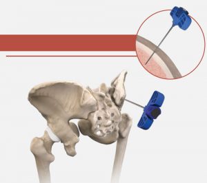
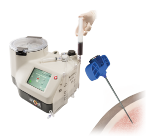
Images: Arthrex
With this new method, stem cells are taken from the iliac crest and immediately processed with a special device. Compared to conventional ACP autologous blood therapy, only very specific growth factors are released and concentrated from the blood, which are particularly beneficial for healing. These are then injected into the bone marrow edema.
Surgical Technique for Bone Marrow Edema – Subchondroplasty
A subchondroplasty in the presence of transient bone marrow edema can shorten the course of the disease.
Subchondroplasty involves filling the bone marrow edema with a special liquid, which on the one hand removes the bone marrow edema and on the other hand supports the adjacent cartilage covering.
Evaluation
The prognosis for traumatic and metabolic bone marrow edema is good with appropriate therapy. A connection between previous traumatic bone marrow edema and secondary cartilage damage is being discussed.