Fracture Malunion and Pseudarthroses
Any fracture healing that deviates from the anatomical norm is referred to as malunion. This can lead to pseudarthrosis. Not all malunions cause a problem. It depends on whether it is a malunion of an articular fracture. The extent of the deviation is relevant, as is the anatomical location.
Each case must be individually evaluated. The therapy decision depends on the given anatomical conditions and the needs (sports, work) of the affected person.
The corrective procedure on the bone is called corrective osteotomy. Corrective osteotomies are necessary to correct axial malalignments, shortenings, and rotational deformities of a bone.
Malalignments of the bone or joint can occur as a result of a malunited fracture or be caused by arthrosis. The newly occurring malalignment leads to excessive stress on the adjacent joints, which in turn causes pain and premature wear.
Locations of Malunions
- Clavicle
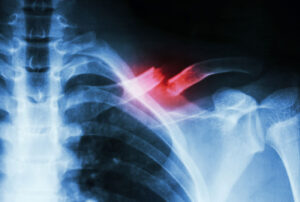 Shortenings and axial deviations, in addition to a cosmetic deficit, lead to excessive stress on the shoulder joint. This can result in pain and chronic complaints. Depending on the malalignment, it can be corrected directly, or the defect in the clavicle can be bridged with a bone graft to restore the correct anatomy. A titanium plate holds the new, correct position in place.
- Malunited Wrist Fracture
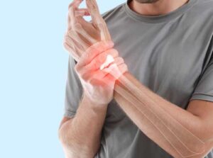 If a wrist fracture heals in the wrong position, this usually leads to mobility issues and causes pain, which can also arise secondarily from premature wear and tear (arthrosis). To correct the malposition, the bone is cut directly at the site of the malposition, an adapted bone graft is inserted, and the position is maintained with a titanium plate. A cast is not necessary after the operation; instead, an anatomically molded removable orthosis is custom-made, allowing early commencement of physiotherapy and movement exercises.
- Malunited metacarpal or finger bones
 Fractures of the fingers and metacarpal bones pose a particular problem, as a malunited finger ray impairs the entire hand function.
Especially rotational deformities have a severe impact, as they significantly disrupt finger closure and impair proper gripping function.
For correction, the bone is cut at the site of the malposition and brought into the correct position. A bone graft is not always necessary for the metacarpals and fingers. Often, the correction can simply be held in position with a titanium plate.
- Periknee Osteotomies for Bow Legs or Knock Knees
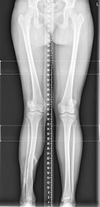 Malalignments of the knee joint can occur after fractures, but also due to arthrosis. An increasing malalignment of the knee joint into a bow-legged or knock-kneed position leads to a progressive shift of the mechanical leg axis, which in turn causes the knee joint to be used very unilaterally and thus to wear out prematurely.
To prevent cartilage damage and early arthrosis, as well as to alleviate pain, a leg axis correction is performed in specific cases. Here, the bone near the knee joint is wedged open at the apex of the deformity and fixed with a titanium plate, thereby slightly overcorrecting the mechanical leg axis to the opposite side. This slight overcorrection relieves the damaged side of the knee.
Pseudarthrosis
A malunited fracture is referred to as pseudarthrosis. This involves the formation of a “false joint” instead of stable bone tissue.
Common Locations of Pseudarthroses
- Clavicle
- Scaphoid Bone
- Ribs
- Metacarpal Bones
- Metatarsal Bones
- Humerus
- Ulna and Radius
- Tibia
A pseudarthrosis can, in principle, develop in any bone and is defined as such when bony healing fails to occur for longer than 6 months in adults.
For the pediatric and adolescent skeleton, shorter periods apply, depending on the respective age.
In pseudarthrosis, pathological mobility occurs. This means that joint-like movements occur in the bone that are not anatomically intended. This leads to pain on the one hand, and the instability of the bone causes discomfort on the other.
There are several types of pseudarthrosis; broadly, they can be divided into hypertrophic and atrophic pseudarthroses.
Cause of Pseudarthrosis
The cause is insufficient blood supply (hypoperfusion) to the fractured area, which prevents bony healing.
This can have several causes:
- Instability
- Insufficient implant
- Incorrectly placed implant
- Incorrectly indicated cast treatment
- Insufficient immobilization or premature loading of the fracture
- Smoking and/or nicotine consumption
- Internal medicine causes, e.g., peripheral arterial occlusive disease (PAOD)
Symptoms of Pseudarthrosis
The symptoms appear gradually
- Increasing pain in the affected area
- Swelling & possibly redness
- Possibly secondary infection of the pseudarthrosis with fever
Pseudarthrosis is diagnosed, in addition to clinical examination, by X-ray examination and computed tomography (CT). Magnetic resonance imaging (MRI) may be required.
Therapy for Pseudarthrosis
The goal of conservative and surgical therapy is to remove the pseudarthrosis. As conservative measures, there are shockwave therapy, immobilization, and splint treatment. If these measures do not lead to healing, a surgical intervention must be performed to achieve bone fracture healing.
The false joint, which consists of a fibrous structure, must be removed, and the defect, if necessary, replaced with autologous bone.
The bone is harvested from the iliac crest using a minimally invasive technique through an approximately 3 cm skin incision, which results in no mechanical restriction of the pelvis.
The affected bone is then firmly fixed in place with a titanium or steel plate so that bony healing can proceed undisturbed. Depending on the location and size of the bone, other implants can also be used for fixation (screws, nails).
For certain indications, a vascularized bone graft is necessary. This is a bone graft that is harvested with a vascular attachment. The micro-surgical connection of the bone segment to the vascular network significantly improves the integration of the transplant.

Dr. Mark Schurz
CONTACT